What are the common conditions when you can see abdominal calcification?
- Atherosclerotic abdominal aorta / aneurysm
- Renal, bladder and ureteral stones
- Gallbladder and common duct stones
- Appendicoliths
- Chronic pancreatitis
- Visceral vascular calcifications
- Calcified cysts in liver and spleen
- Calcified TB nodes
- Dermoid Ovary
What are the imaging procedures useful to demonstrate calcifications in the abdomen?
Detection of calcification in the abdomen is usually an incidental finding.
- Plain abdominal radiographs are particularly useful in showing calcifications.
- 10% of gallstones and 90% of kidney stones will be picked up due to radiopacity from calcium.
- Plain films will show almost all urinary stones since 90% of them are radiopaque
- In addition, appendicoliths, pancreatic calcifications and vascular calcifications (AAA, visceral artery aneurysms, atherosclerosis of visceral vessels) will be picked up.
- CT scan
- Renal stone
- AA
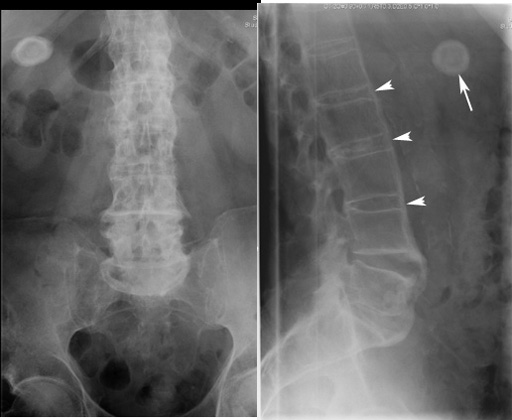
Plain film
Arrow: Gall stone
Arrowheads: Calcification of anterior longitudinal ligament in a patient with ankylosing spondylitis.
Osteophyte L5-S1
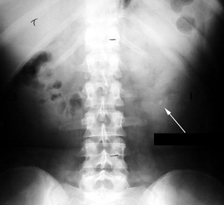
Kidney Stones
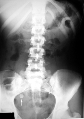
Appendicolith
Findings:
- Arrow showing appendicolith.
- Arrow heads points to ileus.
- Appendicolith may be seen without clinical signs of appendicitis.
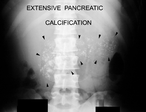
Chronic Pancreatitis
Arrowheads point to extensive pancreatic calcification.
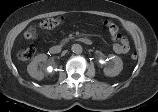
CT
Bilateral Renal Pelvis Calculi
Arrowheads point to soft tissue infiltration secondary to extravasation of urine around both renal pelvis.
Arrows pointing to calculi.
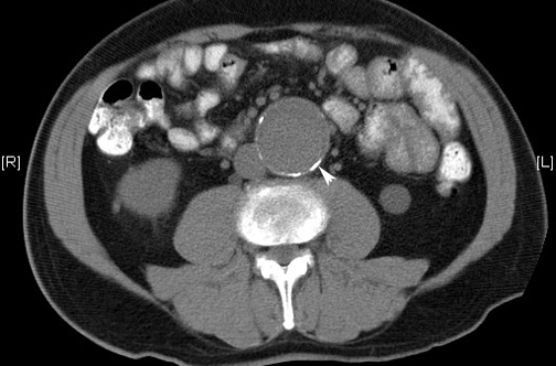
CT
Abdominal Aortic Aneurysm
Calcification of the wall of the aneurysm of aorta