Secondary Bone Tumors / Metastatic Bone Disease
What are the common primaries that metastasize to bones?
- Prostate (most common site from men)
- Breast (most common site from women)
- Lung
- Kidney
- Bladder
- Thyroid
- Lymphomas
Describe the clinical setting and mode of presentation of secondary bone tumors.
- A lesion may be found in an asymtomatic patient with a history of primary tumor.
- Patient with a known primary tumor may present with a painful bone lesion.
- Patient may present with bone pain without known primary cancer and be found to have metastatic bone cancer.
- Patient may present with a pathologic fracture occurring in an area of bone weakened by cancer.
- Patient may present with nerve root or spinal cord compression syndromes.
What are the common bones where metastasis is common?
The sites of metastatic cancer lesions in order of frequency:
- Vertebrae
- Femur
- Pelvis
- Ribs
- Sternum
- Humerus
- Skull
What are the useful imaging modalities to investigate secondary bone tumor?
- Plain Films
- Radioisotope Bone Scan
- CT
- MRI
- PET Scans
- There is no consensus as to the utility of PET scans in the clinical evaluation of bone metastases.
- For some malignancies (i.e., lung and breast cancer), PET scans appear to be more specific than bone scans, although they are possibly less sensitive (especially for osteoblastic foci), and clearly more expensive.
Image Atlas for Secondary Bone Tumors
What are the radiological modes of presentation of secondary bone tumors in plain radiographs?
Plain Films
- Indicates the net result of bone resorption and repair showing osteoblastic, osteolytic and mixed lesions, as well as defining the bone anatomy.
- A skeletal survey (includes lateral skull, cervical spine, anteroposterior (AP) and lateral thoracic and lumbar spine, an AP pelvis and chest radiograph) is used as the primary investigation for bone metastases.
- Lytic lesions must be greater than 1cm in diameter in order to be detected.
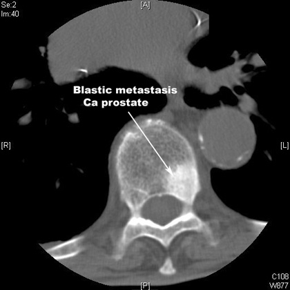
Osteoblastic Lesions
- Result from tumor producing cytokines that activate osteoblasts
- Best detected on bone scan
- Most commonly arises from prostate cancer, but also arise from breast, lung and carcinoid
Blastic Metastasis
- Primary Cancer Prostate
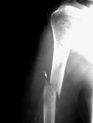
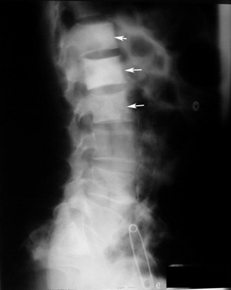
Pathological fracture / Osteolytic metastasis
Lytic lesion of humerus with a pathological fracture.
- Primary Cancer lung.
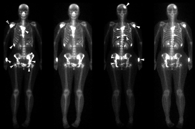
Radioisotope Bone Scan
Excellent screening modality.
Radionuclide bone scanning is the preferred method for evaluating the entire skeleton for the presence of multiple lesions.
- Reflects the metabolic reaction of bone to the disease process, with preferential uptake of the tracer at sites of active bone formation.
- More sensitive than plain films for detecting metastasis.
- Metastases are usually multiple, irregularly distributed foci of tracer that do not correspond to any single anatomic structure.
- The probability that an abnormal scan represents metastatic tumor is directly related to the number of abnormal foci.
- Bone scans are also limited by a lack of specificity, with most false-positive results due to trauma, whether recalled by the patient or not.
- As a result, the diagnosis of metastatic bone disease usually requires radiographic confirmation, especially if the number of lesions is small (fewer than four) and/or limited to the ribs.
Bone Metastasis / Primary Cancer Breast
CT
- Excellent soft tissue image, soft tissue and bony metastases are clearly demonstrated.
- CT most appropriate for diagnosing spinal metastases in locations difficult to assess with bone scan or radiographs (i.e., pelvis/sacrum).
- Also, it is reserved for patients with positive bone scans and negative radiographs in order to clarify pathology.
CT: Osteolytic Lesions
- Result from the tumor production of substances (i.e.PTHrP, Vit D-like steroids) that elicit bone resorption
- Best detected by plain radiology (may not be apparent until > 1cm)
- Most commonly arises from breast, lung, thyroid, renal, melanoma, and gastrointestinal malignancies
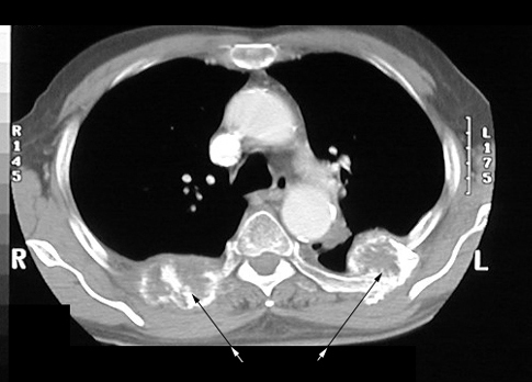
Expansile lytic rib lesions
- Primary Lung Cancer .
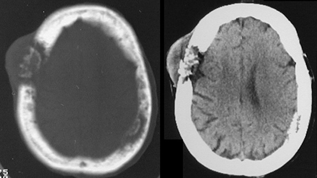
CT
Lytic lesion right parietal bone. Bone destruction with soft tissue mass.
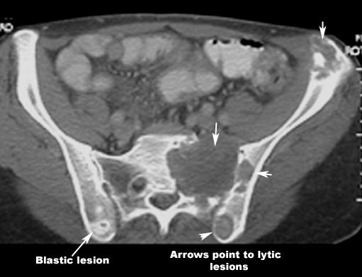
CT: Mixed lesions / Mixture of Osteoblastic and Osteolytic Lesions / Seen in Breast Cancer
Blastic and lytic lesions
- Primary Cancer Breast
- Although whole body MRI has the potential to detect more lesions in the axial skeleton (particularly the spine) than bone scan, it is generally not feasible in most centers due to time constraints.
- Whole body MRI techniques are rapidly advancing, and may replace technetium bone scanning in the future.
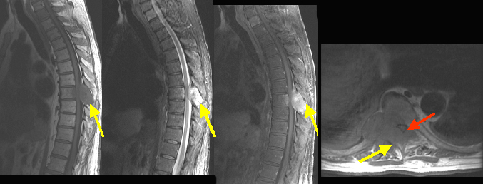
|
Findings: Bony metastasis from renal carcinoma involving the T 8 vertebral body, right pedicle/transverse process and spinous process (arrow in A,B,C,D) with epidural tumor producing marked degree of cord compression (red arrow). |