Primary Bone Tumors
What are the most common primary malignant bone tumors?
- Osteogenic sarcoma
- Ewing's sarcoma
- Fibrosarcoma
- Chondrosarcoma
Primary bone tumors are rare, accounting for less than 1% of all malignant tumors.
What are the useful imaging modalities to investigate primary bone tumors?
- Plain radiographs
- Plain radiographs can often predict the probable histology of a potentially malignant bone lesion.
- MRI
- MRI is the imaging procedure of choice to evaluate primary bone tumors.
- MRI is invaluable in surgical planning as it demonstrates the intraosseus and soft tissue involvement of the tumor and tumor extension.
- MRI is also helpful in evaluating possible malignant degeneration of osteochondromas by allowing accurate measurements of the cartilage cap.
- CT
- CT scans are generally less useful than MRI for assessing primary bone tumors.
- CT is helpful in defining the integrity of the cortex and distribution of calcification.
Image Atlas for Primary Malignant Tumors
What are the imaging findings of primary bone tumor?
- Osteogenic sarcoma
- Destruction of bone
- Sunburst appearance
- Periosteal elevation
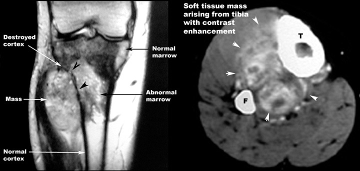
- MR
- Bone scan
- Sclerosis
Osteogenic sarcoma fibula
Destruction of bone with sunburst appearance.
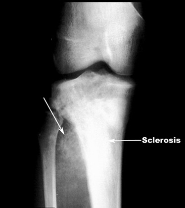
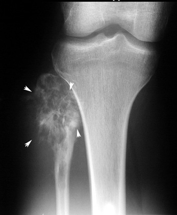
Osteogenic sarcoma Tibia
- Sunburst appearance
- Aggressive periosteal reaction
- Sclerosis
Osteogenic sarcoma tibia
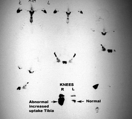
Osteogenic sarcoma tibia
Chondrosarcoma
- Mass with calcification
- On plain radiographs, chondrosarcoma is a fusiform, lucent defect with scalloping of the inner cortex and periosteal reaction.
- Extension into the soft tissue may be present as well as punctate or stippled calcification of the cartilage matrix.
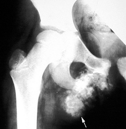
Chondrosarcoma Inferior ramus of pubis
Calcified cartilaginous mass arising from a flat bone in a patient with unfused epiphyses.
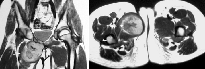
Chondrosarcoma
MR: White arrows : Mass with calcification. Arrowhead is pointing to femur.
-
Ewing's Tumor
- Lytic lesion
- "Onion peel" appearance of periosteal reaction
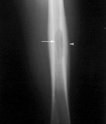
Ewings Sarcoma Femur
- Arrow : Lytic lesion
- Arrowhead: "Onion peel" appearance of periosteal reaction. Layers of periosteum