|
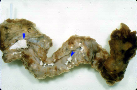
Calcification in pancreas. |
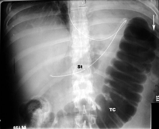
Abdominal x-ray is not diagnostic, but may show:
 |
Acute PancreatitisFindings:
|
 |
Acute PancreatitisCut off sign and Ileus
|
Contrast-enhanced CT of the pancreas is diagnostic and can show:
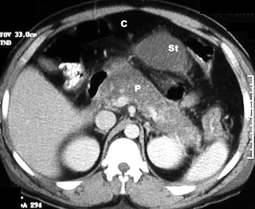 |
Acute PancreatitisCT Findings: Post Contrast
C: Colon
|
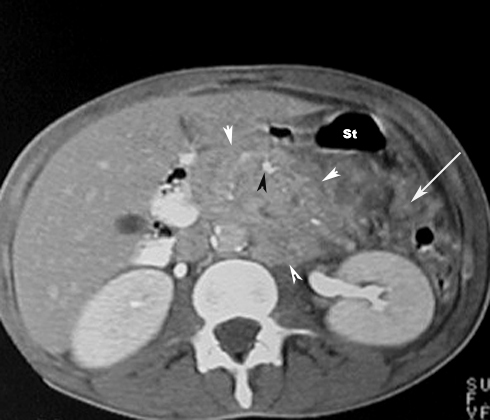 |
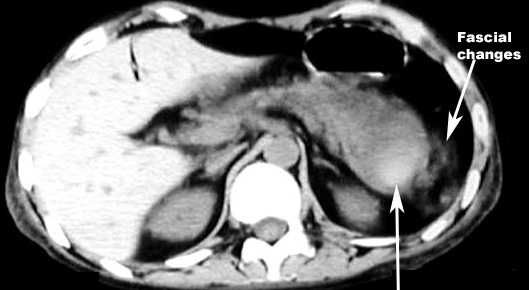
Acute PancreatitisPhlegmon / Inflammatory mass
St: Stomach
|
 |
Acute Pancreatitis / Pancreatic necrosis
St: Stomach
|
 |
Acute Hemorrhagic pancreatitis
|
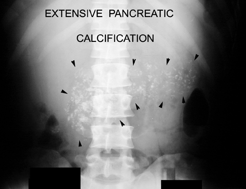 |
Chronic PancreatitisArrowheads point to extensive pancreatic calcification.
|
 |
Pancreatitis with Pseudocyst and calcificationsMass density in pancreas
|