Abdominal Aortic Aneurysm
Pathology
- Atherosclerotic changes leads to aneurysmal dilatation of abdominal aorta
- Calcification of wall can occur
- Complications:
- Rupture with extravasation of blood chronic or acute
- Erosion of the adjacent vertebral bone can occur
- Thrombus can form in the lumen
- Emboli to distal vessels from atheromatous plaques
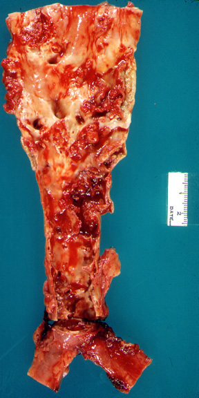
The abdominal aorta was opened along the posterior wall revealing severe atherosclerosis. The anatomical features are obscured by ulcerated plaques and superimposed blood clots.
What are the imaging findings of AAA?
- Widened aortic lumen
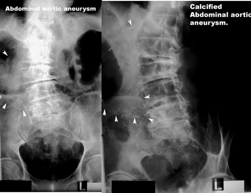
- Plain film:
- Calcification of both walls at the same level with increased diameter
- Displaced calcification of single wall may be due to tortuous undulated aorta
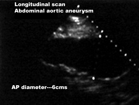
- US: Widened aortic lumen > 3 cm
- Color doppler
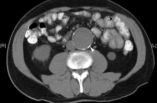
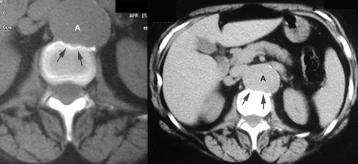
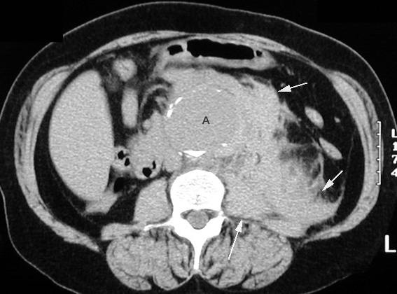
- CT: Dilated aorta
- Calcification of both walls at the same level with increased diameter
- Calcification of the aneurysm wall
- Complications
- Thrombus within aneurysm
- Rupture into
- Retroperitoneum
- Perianeurysmal fibrosis obstructing ureter
- Compression and or erosion of adjacent structures
- Vertebra
- Embolic to distal extremities
- Plain film:
What imaging studies are available for abdominal aortic aneurysm?
- Ultrasound
- Screening modality but only measures aortic diameter.
- For an uncomplicated AAA, ultrasound has a sensitivity and specificity of 97 and 100%.
- For a leaking or ruptured AAA, sensitivity drops to 4% and specificity remains at 100%.
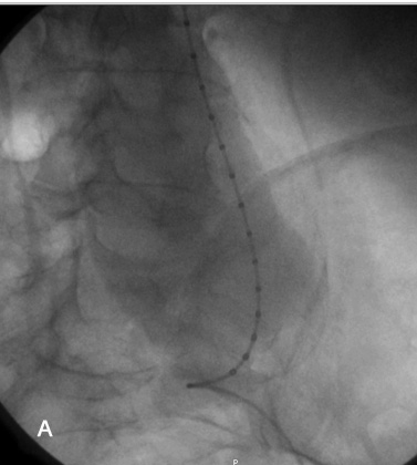
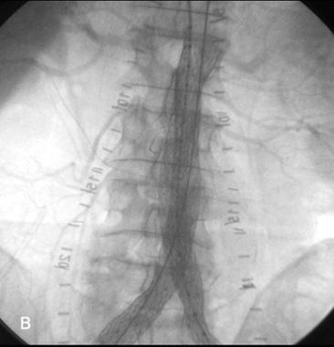
- CT angiogram
- Procedure of choice
- Accurately defines aortic size and extent of aneurysm.
- CT is the first choice in a patient with a classical midline sharp, tearing abdominal pain that radiates to the back, and has a midline pulsatile mass on exam.
- CT has a sensitivity and specificity of 97% and 94%.
- 3D reconstructed images can provide the details necessary for surgical option.
- MRI can provide similar information as a CT angiogram. Difficulty in patient staying still and time of study
- Angiogram: CT angiograms and MRI have replaced the need for angiograms.
- Plain films: Incidental recognition or suspicion of AAA.