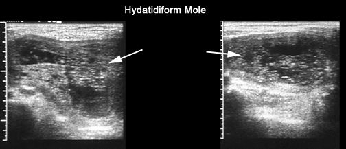
- Uterine cavity filled with multiple sonoluscent areas of varying size and shape, "snowstorm pattern."
- No embryonic or fetal structure.
- No amniotic fluid
- Theca lutein cysts (bilateral, multilocular ovarian cysts > 6 cm in diameter)
- It is possible to have a twin/triplet CHM with a normal fetus in a normal placenta coexisting with the CHM.
- Enlarged placenta containing multi cystic, avascular, sonoluscent spaces, "swiss cheese" appearance.
- Fetal or embryonic tissue is present, may be viable, and is often growth restricted
- Amniotic fluid is present but reduced.
- Increased transverse diameter of gestational sac.
- Theca lutien cysts are absent.
- Diploid/triploid mosaics and twin PHMs are also possible with a normal fetus along with the PHM within the enlarged placenta.