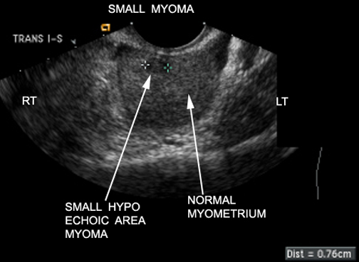
US: Hypoechoic small fibroid
Small anterior wall myoma
MR Coronal Image: Fibroid
Endometrial cavity has some fluid and is seen as a bright stripe.
The fibroids are all hypointense.
Bladder with urine / a bright signal.
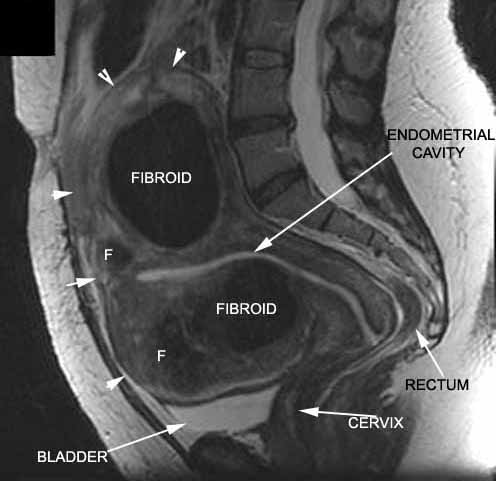
Image Atlas of Fibroid in US, CT and MR
|
US: Hypoechoic small fibroid Small anterior wall myoma |
|
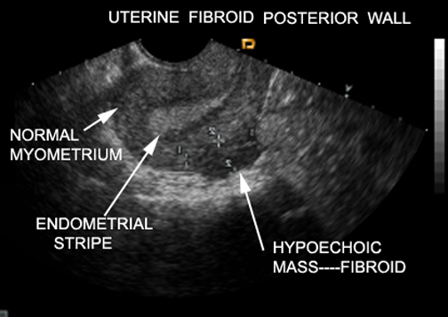
US: Hypoechoic fibroid |
|
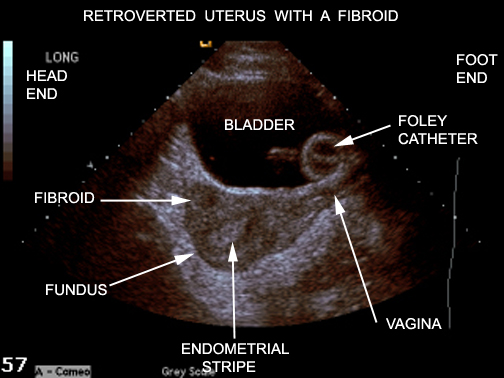
US: Fibroid |
|
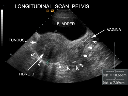
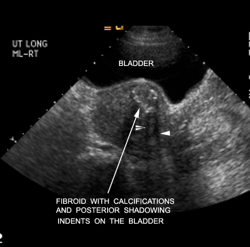
US: Longitudinal scan |
|
US: Fibroid with calcifications |
|
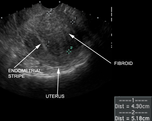
US: Endovaginal uterine fibroid |
|
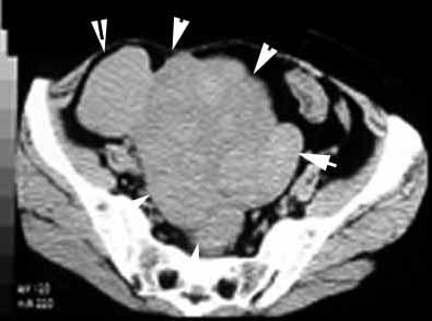
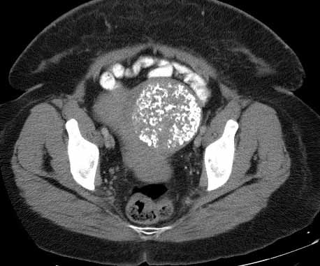
CT: Arrowheads point to enlarged uterus with multiple fibroids. |
|
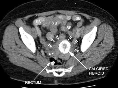
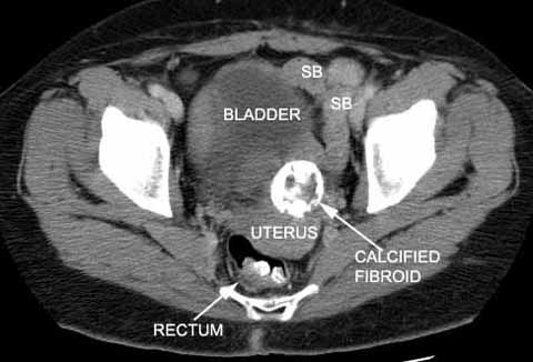
CT Calcified Fibroid. Arrrowheads point to the contour of the uterus |
|
CT Calcified Fibroid. Rectum has contrast from a previous procedure. |
|
CT Calcified Fibroid |
|
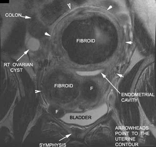
MR Coronal Image: Fibroid Endometrial cavity has some fluid and is seen as a bright stripe. The fibroids are all hypointense. Bladder with urine / a bright signal. |
|
MR Sagittal Image Calcified Fibroid |
60y/o woman who has just noticed an increasing abdominal fullness and minimal abdominal pain and has a large mass on pelvic exam.
Question: What is your initial imaging procedure?
Answer: The procedure of choice is transvaginal ultrasound followed by transabdominal ultrasound, because the larger masses from the fundus cannot be seen on transvaginal ultrasound.
Patient is a 44 year-old G2P2 who presents with heavy bleeding for the past 2 years. She has to change her menstrual pad every hour. She has no abdominal pain. On pelvic exam, her uterus is irregular in shape. She has no adenexal tenderness.
Question: Which imaging prodcedure would you consider?
Answer: Transabdominal ultrasound.