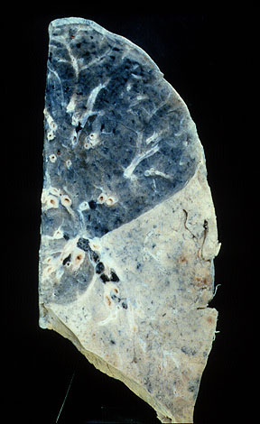
The "gray hepatization" stops abruptly at the fissure.
|
|
The "gray hepatization" stops abruptly at the fissure. |
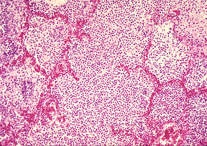
This low power photomicrograph shows many alveolar spaces filled with inflammatory infiltrate. The high power photomicrograph showed the infiltrate to be composed of neutrophiles. Note that the alveolar septa are relatively normal. After complete resolution, the underlying lung architecture is preserved. |
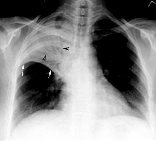 |
What are the x-ray findings of lobar consolidation?
|