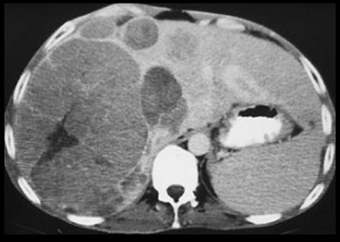
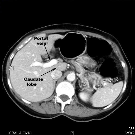
Liver Masses

What are the common liver masses?
Pathology of hepatoma (hepatocellular carcinoma):
What are the anticipated imaging findings of hepatoma?

Normal Liver

  • It is divided into four lobes of unequal size and shape.
  • It is important to understand the complex blood flow (hepatic and portal systems) through the liver. 
  • Parenchyma (Reticulo endothelial cells)  enhances with contrast uniformly with portal vein and hepatic artery branches seen through it.
  • In the superior slices we can see hepatic veins draining into inferior venacava.
  • It is of same density as spleen.
  • Normal biliary ducts are not seen. They are seen only when they are dilated.


CT and MRI are the imaging procedures used in the evaluation of Hepatoma.


CT scan in a patient with Multicentric hepatoma

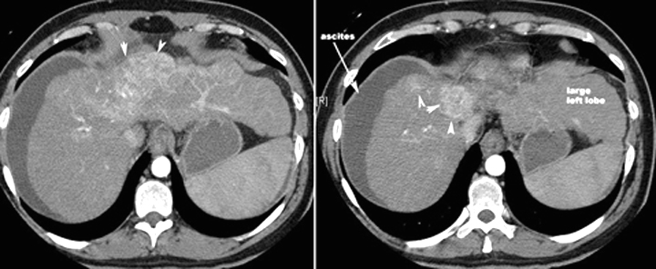
CT scan in another patient with Hepatoma

Arrowheads point to the enhancing mass. Note the lobulated margins of the liver, lower density than spleen and ascites indicating underlying cirrhosis.


Liver metastasis is a common condition presenting as liver mass.
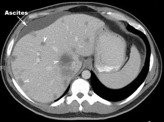
Liver metastasis

Multiple hypo dense lesions seen in the liver with no significant contrast enhancement.

Primary: Colon carcinoma


Liver Cyst

Large cyst left lobe of liver.

  • Oval, Well Defined
  • Imperceptible Or Thin Wall
  • Water Density