Differential Diagnosis: Pelvic Tumor
Case 1
| Ovarian dysgerminoma mimicking appendicitis. 7 year old girl with a history of intermittent fever and right lower quadrant pain for four months. CT was requested to rule out appendicitis. |
|
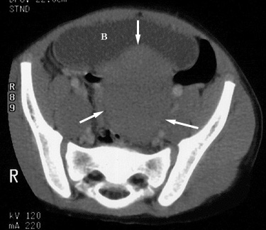
Figure 1. Axial CT image of the pelvis shows a soft tissue mass (arrows) posterior to the bladder (B) |
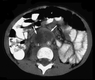 |
Figure 2. Axial CT image of the lower abdomen (same patient as 1.) shows a hypodense mass (arrows) with an enhancing rim anterior to the inferior vena cava (I) and aorta (A) resembling a mesenteric abscess. The inferior vena cava is compressed by the mass. |
| At surgery, the pelvic mass was found to be unresectable; biopsy revealed an ovarian dysgerminoma. |
Return to Differential Diagnosis
Case 2
| Dermoid tumor mimicking appendicitis. 11 year old female presented with severe pelvic pain. Right lower quadrant and pelvic ultrasound, shown below, were requested to evaluate for appendicitis and ovarian torsion. MR imaging, also shown below, was subsequently obtained. |
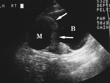 |
Figure 1. Longitudinal transabdominal sonogram shows a thick-walled (arrows) cystic right adnexal mass (M) superior to the urinary bladder (B). |
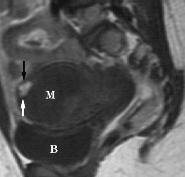 |
Figure 2. Sagittal T1-weighted MR image reveals the complex right adnexal mass (M) to be of predominantly low and medium signal intensity. A small focus of high signal (arrows) within the mass represents fat and is diagnostic of a dermoid tumor. B = urinary bladder. |
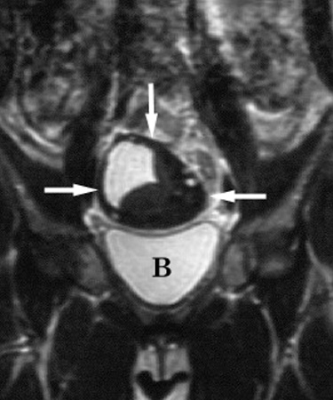 |
Figure 3. Coronal T2-weighted MR image shows areas of both high and low signal intensity within the complex right adnexal mass (arrows). B = urinary bladder. |
| The patient went to surgery, and a right ovarian dermoid tumor was confirmed. |
Return to Differential Diagnosis
Case 3
| Burkitt lymphoma mimicking appendicitis. 4 year old boy with a six week history of pain and tenderness in the right lower quadrant accompanied by fever. A CT examination was performed to evaluate for appendicitis, shown below. |
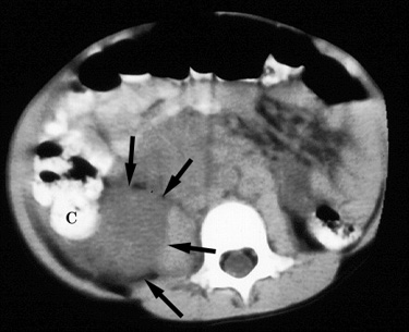 |
Figure 1. Contrast enhanced CT image through the lower abdomen demonstrates a low density paracecal mass (arrows). C = cecum. |
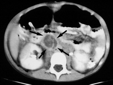 |
Figure 2. CT image slightly superior in location to Figure 1 demonstrates another, separate mass (arrows), anterior to the inferior vena cava, with an enhancing rim and low attenuation central portion, resembling a mesenteric abscess. |
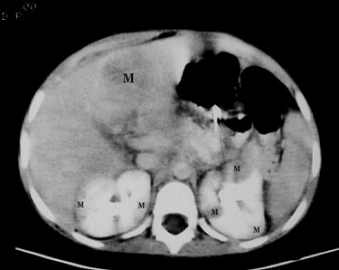 |
Figure 3. CT image slightly superior in location to Figure 2 shows a low density mass (large M) in the left lobe of the liver. Additional low attenuation masses (small M's) are present in the kidneys bilaterally. |
| Biopsy revealed Burkitt lymphoma. The patient was treated with chemotherapy, resulting in regression of the tumor. |
Return to Differential Diagnosis