Atypical Clinical Presentation: Retrocecal Appendix
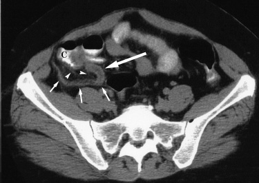
| Nonperforated appendicitis. 16 year old girl with focal pain, right lower quadrant tenderness, and fever for two days. A CT examination was obtained to evaluate for appendicitis, shown below. |
|
An axial CT image in the upper pelvis shows edema of the cecal wall which, along with barium in the cecum (C), contributes to the "arrowhead sign" of appendicitis. A dilated fluid filled appendix (large arrow) is seen with adjacent stranding of retroperitoneal fat (arrowheads). The appendix follows a retrocecal course (small arrows). |
| At surgery a nonperforated inflamed appendix was removed. |
Return to Atypical Clinical Presentation (A)