Plain Film and Barium Enema Findings in Appendicitis
| Perforated appendicitis with multiple
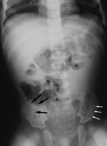
appendicoliths and ileus. 1 year old girl presented to the emergency room with abdominal distention and fever. A plain abdominal radiograph was obtained, shown below. |
|
Supine anteroposterior abdominal radiograph demonstrates several calcified appendicoliths (black arrows) in the right lower quadrant. Note loss of visualization of the properitoneal fat on the right (compare with normal appearance on the left, white arrows), consistent with an adjacent inflammatory process. There are several mildly dilated small bowel loops in the left upper and right lower quadrants (B). |
| An ultrasound was requested to evaluate for appendicitis, shown in Ultrasound Findings in Appendicitis. |
Return to Atypical Clinical Presentation (C)
Return to Plain Film Findings
| Pericecal appendiceal abscess. 17 year old male with history of right lower quadrant pain for one week. A barium enema, shown below, was performed to evaluate for appendicitis. |
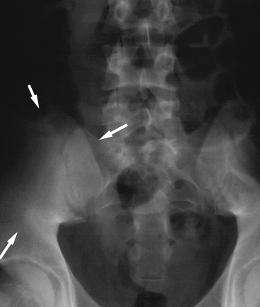 |
Figure 1. Scout film of the abdomen shows a paucity of bowel gas in the right lower quadrant (arrows)consistent with a soft tissue mass. |
 |
Figure 2. Barium enema demonstrates mass effect on the base of the cecum (C), resulting in a "coiled-spring" appearance. The latter can be seen with peri-appendiceal or peri-cecal abscess. The abscess also produces mass effect on the distal ileum (I). |
| At surgery, a perforated appendix and adjacent abscess were found. |
Return to Plain Film Findings
Return to Barium Enema Findings
| Perforated appendicitis with pericecal
abscess causing small bowel obstruction . 2 year old boy presented with signs and symptoms of small bowel obstruction for one day. An abdominal radiograph, shown below, was obtained. |
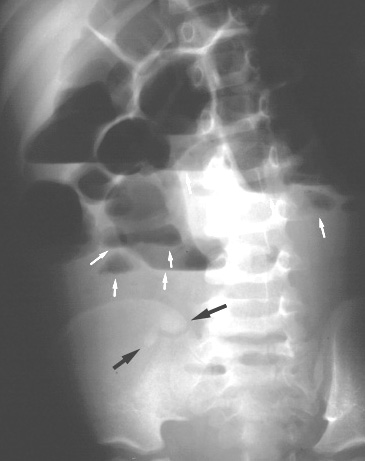 |
Upright abdominal radiograph shows dilated small
bowel loops containing air fluid levels (small white
arrows), consistent with small bowel obstruction. Two
calcified appendicoliths (large black arrows) are seen in
the right lower quadrant which is devoid of bowel gas due
to the presence of an abscess. Additional findings include scoliosis of the lumbar spine with convexity to the left. |
| Surgery confirmed the findings of perforated appendicitis and adjacent abscess. |
Return to Plain Film Findings
| Perforated appendicitis with a mildly dilated
appendix. 14 year old girl presented to the emergency room with two day history of fever, right lower quadrant pain, and emesis. Laboratory evaluation showed leukocytosis. Plain abdominal radiograph was obtained, shown below. CT was also performed to evaluate for appendicitis, discussed in CT Findings section. |
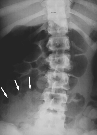 |
Abdominal radiograph demonstrates a mild, diffuse ileus, paucity of right lower quadrant bowel gas (arrows), and scoliosis with leftward convexity. |
| A subsequent CT examination revealed a non-dilated appendix with peri-appendiceal and regional retroperitoneal fluid, shown in CT Findings section. At laparoscopic surgery, there was perforated appendicitis and a small peri-appendiceal abscess. |
Return to Plain Film Findings
| Perforated appendicitis. 4 year old male with two day history of fever, abdominal pain and vomiting. Supine anteroposterior and cross-table lateral radiographs, shown below, were the only diagnostic studies obtained. |
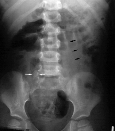 |
Figure 1. Supine anteroposterior abdominal radiograph shows a calcified appendicolith (white arrows) in the right lower quadrant. The bowel gas pattern is nonspecific, with mildly dilated, air-filled small bowel loops in the right upper quadrant. The left psoas muscle margin (black arrows) is normal and distinct; the right psoas muscle margin is not seen. |
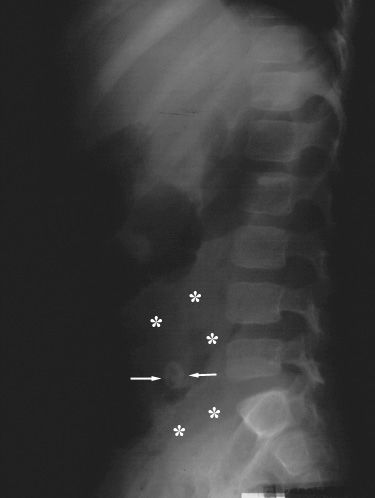 |
Figure 2. Supine cross-table lateral abdominal radiograph shows the calcified appendicolith (arrows) and a paucity of regional bowel gas (*). |
| The patient underwent an emergent appendectomy, and perforated appendicitis was discovered. |
Return to Plain Film Findings
| Perforated appendicitis. 6 year old male with one day history of right lower quadrant pain and fever. On physical examination, the patient's pain was reproduced and severe with only mild palpation. A supine abdominal radiograph, shown below, was the only diagnostic study obtained. |
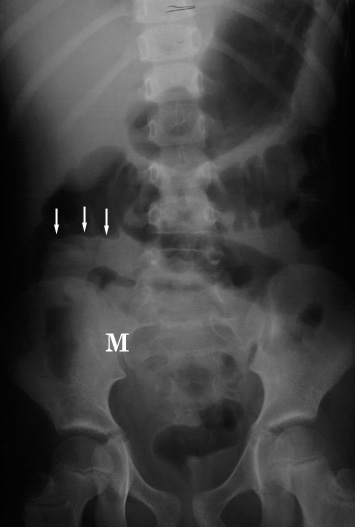 |
Supine abdominal radiograph demonstrates a paucity of bowel gas in the right lower quadrant with a suggested underlying soft tissue mass (M). The colonic gas column abruptly terminates ("colon cut-off" sign) proximal to the hepatic flexure (arrows). Note mild scoliosis, concave to the right. |
| The clinical and plain radiographic findings were highly suggestive of appendicitis, and the patient underwent an emergent appendectomy. Perforated appendicitis with abscess formation was discovered intraoperatively. |
Return to Plain Film Findings