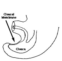
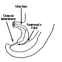
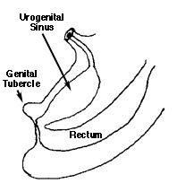
In the early stages of development, there is no separation of the urinary and alimentary tracts. A common chamber, known as the cloaca, forms in the caudal region of the fetus. At the caudal end of the cloaca, ectoderm lies directly over endoderm forming the thin cloacal membrane. As development progresses, a septum forms (Toureux's fold) dividing the hind gut from an anterior chamber, the urogenital sinus. This septum extends in a caudal direction. Two tissue folds arise from the lateral sides of the cloaca (Rathke's plicae). These folds move medially toward each other to complete the separation of the hind gut from the urogenital sinus. Tourneux's folds and Rathke's plicae together form the uro-rectal septum.
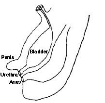
At 10 weeks gestation, the bladder is a cylinder. The cranial portion of the cylinder tapers to become the vesico-allantoic canal. By 12 weeks, the vesico-allantoic canal closes completely leaving the median umbilical ligament. What happens if this tract fails to close?
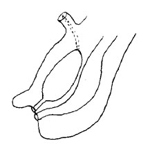
See an axial plane movie of normal bladder development.
To next page of Normal Bladder Development
Return to G/U Development Home Page
©David A. Hatch, M.D., 1996