Head Trauma
Q1: What are the common symptoms of head injury?
- Symptoms of head injury can vary greatly and depend on the type, location, and extent of injury.
- The most common symptoms are headache, nausea, vomiting, altered mental status, and/or loss of consciousness.
- Symptoms are neither sensitive nor specific for detecting an intra cranial abnormality.
Q2: What are the various radiological manifestations of head injury?
- Skull Fractures :
- Linear fracture
- Depressed fracture
- Basilar fracture
- Epidural hematoma
- Subdural Hematoma
- Diffuse Axonal Injury
- Contusions (cerebral hemorrhages)
- Pneumocephalus
 |
Skull Fractrures |
Q3: What are the available procedures and their utility in the evaluation of head trauma?
- In moderate to severe head trauma cases, non-contrast- enhanced CT scanning is the most useful initial imaging technique.
- CT will detect acute hemorrhage, cerebral swelling, evidence of elevated intracranial pressure and pneumocephalus..
- Using the bone window, it will also detect skull fractures, eliminating the need for additional plain skull films.
- CT scans are rapid, readily available at most institutions, and accessible for unstable patients.
- While MRI has limited applications in the immediate posttraumatic period, it is more sensitive than CT in detecting sub acute non hemorrhagic lesions, axonal shear injury, and brainstem or posterior fossa lesions.
- Skull x-rays are rarely necessary and have been replaced by CT.
- Whether to perform imaging studies in minor head trauma cases is a controversial issue.
Case 1:
A 50 year old previously healthy male one day after a motor vehicle accident, presents to his physician's office, just to make sure that everything is okay. Yesterday, the patient was in the driver's seat stopped at a red light when he was rear-ended by a truck at approximately 35-40 mph. He was wearing his seatbelt which prevented him from hitting the steering wheel or dashboard, but the back of his head did snap back into the headrest. The patient says he feels fine but was urged by his wife to get a checkup anyway. He denies any symptoms. The neurological examination is normal.
Is there a need for imaging studies in this case? Why or why not? What are the medico legal issues of head trauma?
- The emergency management of minor head trauma is very controversial and revolves around whether to perform imaging studies or not.
- Initial CT scanning (and observation when the scan is normal) of all minor head trauma patients would allow early detection of delayed complications or deterioration.
- However, this is very costly since the risk of intra cranial abnormalities or delayed complications is minimal.
- CT scan should be considered when there are focal neurological findings, persistent headache, vomiting, and age >60.
- In asymptomatic patients, with normal neurological exam, without other injuries or loss of consciousness, neuroimaging is not indicated and the patient can be observed at home safely.
- The controversy continues for now and physicians continue to make decisions on a case-by-case basis.
Case 2:
A 58 year old alcoholic male was brought to the hospital after a fall down a flight of stairs. His neighbor reported he fell head first but denied any loss of consciousness. The patient was disoriented. The neurological exam was significant for left hemiparesis, a left Babinski sign, and a left homonymous hemianopsia.
What is your working diagnosis?
Sub dural hematoma
Which imaging procedure will you order?
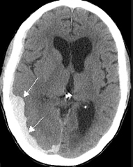
Non contrast CT head. Head CT showed a large crescenteric right-sided acute subdural hematoma.
|
|
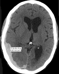 NNNoncontrast CT NNNoncontrast CT
CT density of blood is 74 HU consitant with acute blood |
|
Diagnosis: Acute Subdural Hematoma
- Head CT showed a large crescenteric right-sided acute subdural hematoma.
- Acute subdural hematoma covering the right cerebral hemisphere (arrows), more prominent posteriorly.
|
Case 3:
A 35 year old male was involved in a motor vehicle accident and was brought to the hospital by ambulance complaining of a severe headache. He was reported to be disoriented and combative at the scene . En route to the ER, the patient's mental status improved and he was more cooperative. However, upon arrival in the ER the patient's mental status worsened. He had no memory of the accident. On physical examination, the patient had swelling around the left temporal region as well as left periorbital ecchymosis.
What is your working diagnosis?
Epidural hematoma
Which imaging procedure will you order?
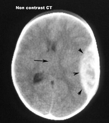
Non contrast CT. CT showed a left-sided well-defined biconvex extra-axial mass consistent with an acute epidural hematoma. Bone windows showed a linear fracture of the skull.
|
Epidural Hematoma
- CT showed a left-sided well-defined biconvex extra-axial mass consistent with an acute epidural hematoma.
- Arrowheads point to the epidural hematoma, which is a collection of blood between the skull and the dura.
- It typically has a biconvex margin towards the brain surface.
- Arrow points to shift of midline to the right.
- Bone windows showed a linear fracture skull.
|
Case 4:
A 25 year old male was brought to the emergency room by ambulance after sustaining a blow to the head with a baseball bat during an altercation between himself and another person. The patient lost consciousness immediately but then regained consciousness within several minutes and was disoriented until arrival at the hospital. Currently he is alert and oriented and complaining of a bilateral headache, worse on left. On physical exam a tender swelling around the left forehead was seen. No focal neurological deficits were present and the optic discs were flat.
What is your working diagnosis?
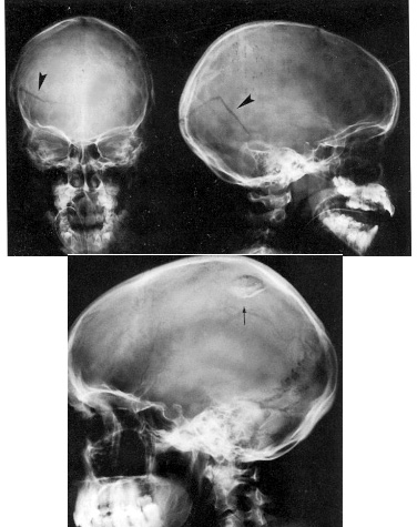
Skull fracture
Which imaging procedure will you order?
CT scan which confirmed the left frontal skull fracture (bone window) and ruled out underlying parenchymal injury (brain window).
What is meant by the terms bone and brain window in CT?
- Bone window focuses on bony structure. Brain window focuses on soft tissues.
- When a skull fracture is suspected bone windows should be obtained.