Normal Development of the Testis and Scrotum
David A. Hatch, M.D.
Loyola University Stritch School of Medicine
At 3 weeks of development, the germ cells migrate from the yolk sac
to the genital ridge. From the 4th to the 8th week, in male embryos with
a normal sex determining region on the short arm of the Y chromosome, the
germ cells coalesce to form the primordial testis. Under the influence
of human chorionic gonadotropin , the Leydig cells of the
developing testis begin to secrete testosterone.
At about 9 weeks of development, the labioscrotal swellings fuse
to form the scrotum (see movie). Testosterone
also induces development of the mesonephric (Wolfian) duct to form
the epididymis, vas deferens and seminal vesicles.
During this stage of development, the testis moves from the genital
ridge across the pelvis to lie at the internal inguinal ring.
The processus vaginalis appears at about 13 weeks of development
as an outpouching of the parietal peritoneum. This developing tunnel moves
medial and caudal between the internal and external abdominal oblique muscles
and into the scrotum. The testis stays at the opening of the processus
vaginalis, the internal inguinal ring, for 10 to 12 weeks. This herniation
of the patent processus is at least partially dependent on the abdominal
wall musculature to generate an elevated intra-abdominal pressure. If the
abdominal muscles cannot increase intra-abdominal pressure, the patent
processus vaginalis may not progress through the inguinal canal and the
testis may not descend into the scrotum
At 26 to 36 weeks of development the epididymis precedes the testis
into the processus vaginalis. These structures descend into the scrotum
and become fused with the posterior layers of the scrotum, providing an
anchor which prevents the testis from rotating. At 37 to 40 weeks (full
term), the processus vaginalis closes, eliminating any communication between
the peritoneum and the inguinal canal or scrotum.
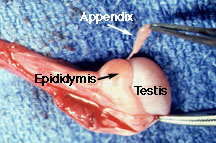
| As the mesonephric duct develops into the epididymis, a
proximal remnant (or more than one remnant) may persist as a small appendage,
the appendix epididymis. This tissue is most often attached to the caput
epididymis (the most proximal and cephalad portion). Occasionally, such
an appendix can twist and become inflamed. See
a case history.
Simultaneously, the paramesonephric structures (Müllerian) regress
under the influence of Müllerian inhibiting substance secreted
by the Sertoli cells of the developing testis. The most proximal
remnant of the Müllerian duct persists as the appendix testis. |
|
At any one of these developmental stages, anomalies can occur which result
in problems after birth.
See abnormal testis/scrotal development.
Return to G/U Development Home Page.
©David A. Hatch, M.D., 1996